ESD (Endoscopic Submucosal Dissection)
Endoscopic Submucosal Dissection (ESD) is an advanced minimally invasive endoscopic procedure used to remove selected abnormal or early-stage lesions from the lining of the digestive tract. It allows the lesion to be removed carefully and, in many suitable cases, in a single piece while preserving the surrounding healthy tissue.
1. Patient Evaluation
Before ESD, the patient undergoes a detailed clinical assessment. The location, size, appearance, and characteristics of the lesion are evaluated to determine whether ESD is an appropriate treatment option.
2. Pre-Procedure Assessment
Depending on the patient's condition, investigations such as blood tests, endoscopic assessment, biopsy, or imaging may be recommended. The patient's medical history, medications, and overall health are also reviewed before the procedure.
3. Endoscopic Examination
A specialized endoscope is carefully introduced into the digestive tract to provide a clear view of the lesion. The lesion is examined in detail to determine its boundaries and depth of involvement.
4. Lesion Marking
The margins of the lesion are carefully identified and marked endoscopically. This helps the specialist define the area that needs to be removed while minimizing unnecessary damage to surrounding tissue.
5. Submucosal Injection
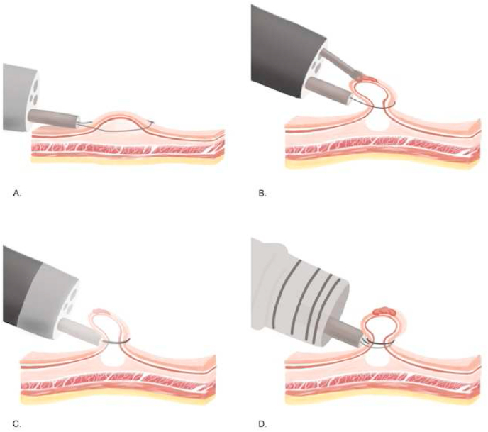
A suitable solution is injected beneath the lesion to lift the affected tissue away from the deeper layers. This creates a safer working space for precise dissection.
6. Mucosal Incision
A controlled incision is made around the marked lesion using specialized endoscopic instruments. The procedure is performed carefully to maintain a clear view and protect the deeper layers of the digestive tract.
7. Submucosal Dissection
The specialist carefully dissects beneath the lesion through the submucosal layer. The abnormal tissue is gradually separated from the underlying layers with precision.
8. Complete Lesion Removal
The objective is to remove the targeted lesion completely, often as a single specimen when technically appropriate. Removing the lesion in one piece can help provide an accurate pathological assessment of the margins and tissue characteristics.
9. Specimen Assessment
The removed tissue is sent for histopathological examination. Laboratory analysis helps determine the exact nature of the lesion, depth of invasion, and whether the removal margins are clear.
10. Post-Procedure Monitoring
After ESD, the patient is monitored for recovery and possible complications. Instructions regarding diet, medications, activity, and follow-up are provided according to the individual case.
11. Follow-Up Care
Follow-up endoscopy or additional investigations may be recommended depending on the pathology results and the characteristics of the lesion. Continued monitoring helps assess healing and detect any recurrence at an early stage.
Benefits of ESD
ESD can provide precise removal of selected superficial gastrointestinal lesions while avoiding conventional open surgery in appropriate cases. It may also allow the removed tissue to be examined as a complete specimen, providing valuable information for accurate diagnosis and further treatment planning.
Comprehensive ESD Care
Our ESD service includes appropriate patient assessment, detailed endoscopic evaluation, advanced lesion removal, pathological examination, post-procedure monitoring, and follow-up care. ESD is selected on an individual basis after careful evaluation of the lesion and the patient's overall condition.