Foreign Body Removal by Endoscopy:
Introduction
Foreign body removal by endoscopy is a minimally invasive medical procedure used to remove swallowed objects from the gastrointestinal tract. These objects may become lodged in the esophagus, stomach, or duodenum and can cause pain, obstruction, bleeding, or tissue damage. Endoscopy allows a trained medical professional to visualize the foreign body and remove it using specialized instruments.
Early diagnosis and appropriate treatment are important to reduce the risk of complications and improve patient outcomes.
What Is Foreign Body Removal by Endoscopy?
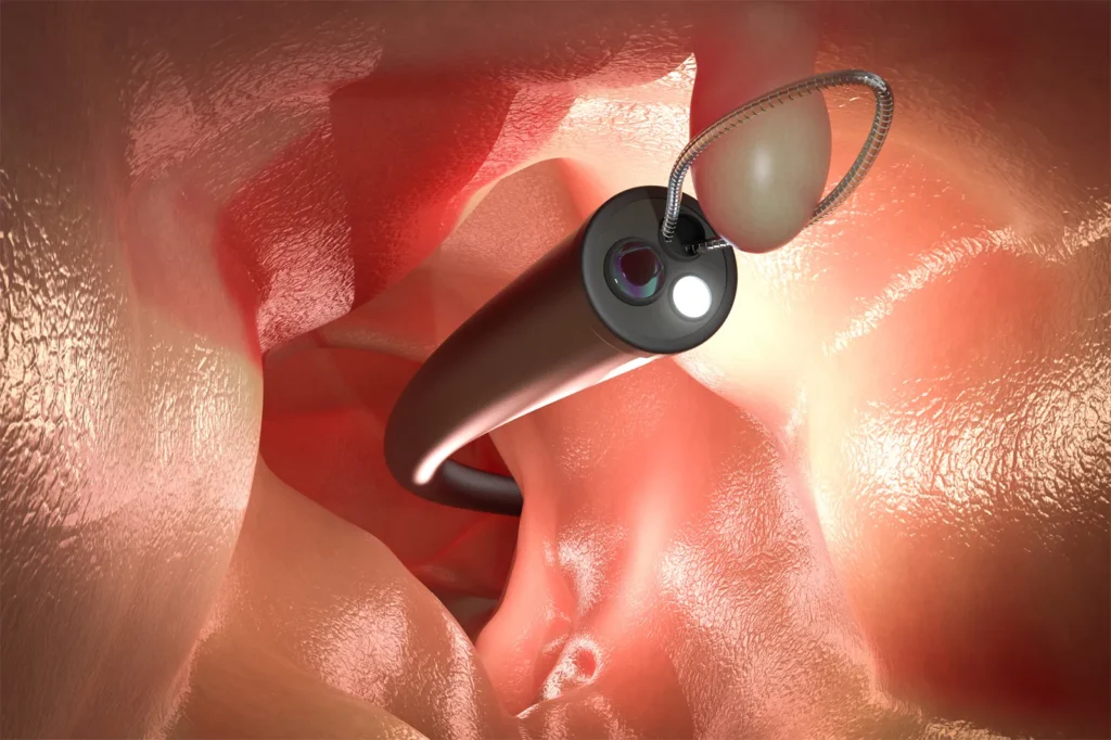
Foreign body removal by endoscopy is a procedure in which a flexible tube called an endoscope is introduced into the gastrointestinal tract. The endoscope contains a camera and light source that help the doctor examine the digestive tract and locate the foreign object.
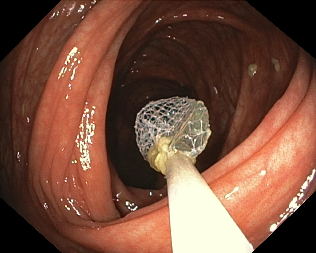
Special instruments, such as retrieval forceps, snares, baskets, or retrieval nets, may be passed through the endoscope to grasp and remove the object safely.
The procedure is commonly performed by a gastroenterologist or another appropriately trained specialist.
Common Indications for Foreign Body Removal
Endoscopic foreign body removal may be required in the following situations:
- Swallowed coins or metallic objects.
- Impacted food boluses.
- Fish bones or chicken bones.
- Swallowed dentures or dental prostheses.
- Sharp objects such as pins, needles, or toothpicks.
- Button batteries lodged in the esophagus.
- Multiple swallowed magnets.
- Foreign bodies causing gastrointestinal obstruction.
- Objects that remain lodged in the stomach or upper gastrointestinal tract and require removal.
The urgency of treatment depends on the type of object, its location, the duration of ingestion, and the patient's symptoms.
Types of Foreign Bodies
Foreign bodies vary in size, shape, and potential to cause injury.
1. Blunt objects: Coins, small toys, and other smooth objects may become lodged in the esophagus or pass through the digestive tract.
2. Sharp objects: Pins, needles, bones, and other pointed objects can injure the gastrointestinal lining and may cause perforation.
3. Food bolus: Food may become impacted in the esophagus, causing difficulty swallowing or inability to swallow saliva.
4. Button batteries: These can rapidly damage surrounding tissues, particularly when lodged in the esophagus, and require emergency assessment.
5. Magnets: Multiple magnets, or a magnet swallowed with another metallic object, can attract across intestinal walls and cause serious injury.
Types of Endoscopy Used
Esophagoscopy
Esophagoscopy is used to examine the esophagus and remove foreign bodies lodged in this part of the digestive tract.
Upper Gastrointestinal Endoscopy (EGD)
Upper gastrointestinal endoscopy allows examination of the esophagus, stomach, and duodenum. It is commonly used to retrieve foreign bodies from the upper gastrointestinal tract.
Colonoscopy
In selected cases, colonoscopy may be considered to retrieve a foreign body located in the colon, depending on its position and the patient's condition.
Bronchoscopy
Bronchoscopy is used to remove foreign bodies from the respiratory tract. It is different from gastrointestinal endoscopy and is indicated when an object enters the airway.
Preparation Before the Procedure
Proper preparation helps improve patient safety and procedural success.
- Medical history: The doctor reviews the patient's medical history, symptoms, medications, and relevant medical conditions.
- Identification of the object: Information about the type, size, shape, number, and time of ingestion helps determine the appropriate treatment.
- Physical examination: The patient is assessed for airway compromise, difficulty swallowing, abdominal tenderness, bleeding, and other warning signs.
- Imaging investigations: X-rays or CT scans may be performed to identify the location of the foreign body and assess possible complications.
- Laboratory tests: Blood tests may be required depending on the patient's clinical condition and the planned intervention.
- Informed consent: The procedure, potential benefits, risks, and alternatives are explained to the patient or their legal guardian.
- Anesthesia assessment: The need for sedation, general anesthesia, and airway protection is evaluated.
- Fasting assessment: The patient's last oral intake is reviewed. Urgent foreign body removal should not be delayed solely to complete routine fasting when emergency intervention is necessary.
Equipment Used for Endoscopic Foreign Body Removal
The equipment selected depends on the nature and location of the foreign body.
Commonly used equipment includes:
- Flexible gastrointestinal endoscope.
- Video monitor and light source.
- Grasping or retrieval forceps.
- Retrieval basket.
- Polypectomy snare.
- Retrieval net.
- Protective cap or hood.
- Overtube, when indicated.
- Suction and irrigation equipment.
- Bite block.
- Oxygen supply and monitoring equipment.
- Emergency resuscitation equipment.
Specialized retrieval accessories help the endoscopist grasp, secure, and withdraw the foreign body while minimizing injury to surrounding tissues.
Step-by-Step Procedure for Foreign Body Removal by Endoscopy
Step 1: Patient Assessment
The patient is assessed to determine the urgency of removal, the location of the foreign body, and the risk of complications.
Step 2: Patient Positioning
The patient is positioned appropriately for the planned endoscopic procedure. The left lateral position is commonly used for upper gastrointestinal endoscopy.
Step 3: Monitoring and Anesthesia
Appropriate monitoring is established. Sedation or general anesthesia may be used depending on the patient's condition, aspiration risk, and procedural complexity.
Step 4: Insertion of the Endoscope
The endoscope is introduced through the mouth and advanced under direct visualization toward the suspected location of the foreign body.
Step 5: Identification of the Foreign Body
The endoscopist identifies the object and evaluates its size, shape, orientation, and relationship to the surrounding gastrointestinal lining.
Step 6: Selection of Retrieval Instruments
The appropriate retrieval accessory is selected according to the type of object. Forceps, snares, baskets, or retrieval nets may be used.
Step 7: Grasping the Object
The foreign body is carefully secured using the selected instrument. The technique is chosen to provide a stable grip and reduce the risk of tissue injury.
Step 8: Safe Extraction
The object is withdrawn carefully under endoscopic visualization. Protective devices may be used when appropriate, particularly for sharp objects or objects with irregular edges.
Step 9: Inspection of the Gastrointestinal Tract
After removal, the endoscopist examines the affected area for bleeding, ulceration, lacerations, perforation, or retained fragments.
Step 10: Confirmation and Recovery
Successful retrieval is documented, and the patient is transferred to the recovery area for observation and monitoring.
Possible Complications
Although endoscopic foreign body removal is generally considered safe when appropriately performed, complications can occur.
Potential complications include:
- Bleeding.
- Mucosal abrasions or lacerations.
- Perforation of the gastrointestinal tract.
- Aspiration of stomach contents.
- Respiratory depression associated with sedation.
- Infection in selected cases.
- Migration of the foreign body.
- Incomplete retrieval.
- Need for additional endoscopic intervention or surgery.
The risk of complications depends on the foreign body's characteristics, its location, the duration of impaction, and the patient's overall condition.
Post-Procedure Care
Appropriate monitoring after the procedure helps identify complications early.
- Monitor vital signs and recovery from sedation.
- Assess for chest pain, abdominal pain, breathing difficulty, and bleeding.
- Follow the treating team's instructions regarding oral intake.
- Provide medications when clinically indicated.
- Observe the patient for signs of perforation or other complications.
- Document the outcome of the procedure and any injuries identified.
- Arrange follow-up care when required.
Severe or worsening pain, fever, difficulty breathing, vomiting blood, or black stools require urgent medical assessment.
Nursing Responsibilities
Before the Procedure
- Verify patient identity and the planned procedure.
- Review consent, allergies, medications, and relevant investigations.
- Assess baseline vital signs and fasting status.
- Prepare the endoscope and retrieval accessories.
- Ensure that oxygen, suction, monitoring, and emergency equipment are available.
- Provide patient education and reassurance.
During the Procedure
- Assist the endoscopist as required.
- Maintain appropriate patient positioning.
- Monitor vital signs and oxygen saturation.
- Prepare and handle retrieval instruments.
- Maintain infection prevention and equipment safety.
- Report any deterioration in the patient's condition immediately.
After the Procedure
- Monitor the patient's recovery from sedation.
- Assess for pain, bleeding, respiratory problems, and other complications.
- Follow instructions regarding restarting oral intake.
- Maintain patient safety until adequate recovery.
- Document the procedure and post-procedure observations.
- Explain discharge instructions and warning signs.
When Is Foreign Body Removal an Emergency?
Urgent medical assessment is necessary when a patient has:
- A button battery lodged in the esophagus.
- A sharp object lodged in the gastrointestinal tract.
- Complete esophageal obstruction or inability to swallow saliva.
- Multiple swallowed magnets or a magnet swallowed with a metallic object.
- Severe chest or abdominal pain.
- Signs of gastrointestinal perforation or significant bleeding.
- Breathing difficulty or suspected airway obstruction.
The timing of endoscopy depends on the object and the patient's clinical condition. Emergency cases require prompt assessment by an appropriate medical team.
Frequently Asked Questions
Is foreign body removal by endoscopy painful?
The procedure is commonly performed with sedation or anesthesia to improve patient comfort. Some patients may experience temporary throat discomfort afterward.
How long does the procedure take?
The duration depends on the location, size, and type of foreign body, as well as the complexity of removal.
Is surgery always required to remove a swallowed foreign body?
No. Many foreign bodies in the upper gastrointestinal tract can be removed endoscopically. Surgery may be necessary if endoscopic removal is unsuccessful or complications require surgical treatment.
What happens after the foreign body is removed?
The medical team monitors the patient for complications and determines when oral intake can safely resume. Further treatment may be required if tissue injury is present.
Can all swallowed objects be removed by endoscopy?
No. The suitability of endoscopic removal depends on the object's location, shape, size, and associated complications. Some cases require alternative management or surgery.
Conclusion
Foreign body removal by endoscopy is an important minimally invasive procedure used to retrieve swallowed objects from the gastrointestinal tract. Appropriate patient assessment, timely intervention, suitable retrieval instruments, and careful post-procedure monitoring are essential for safe and effective management.
Early medical evaluation is particularly important when the swallowed object is a button battery, a sharp item, or a foreign body causing obstruction.
Medical Disclaimer: This article is intended for educational purposes only and does not replace professional medical advice, diagnosis, or treatment. Suspected foreign body ingestion requires assessment by a qualified healthcare professional.