Gastrointestinal Stromal Tumor (GIST)
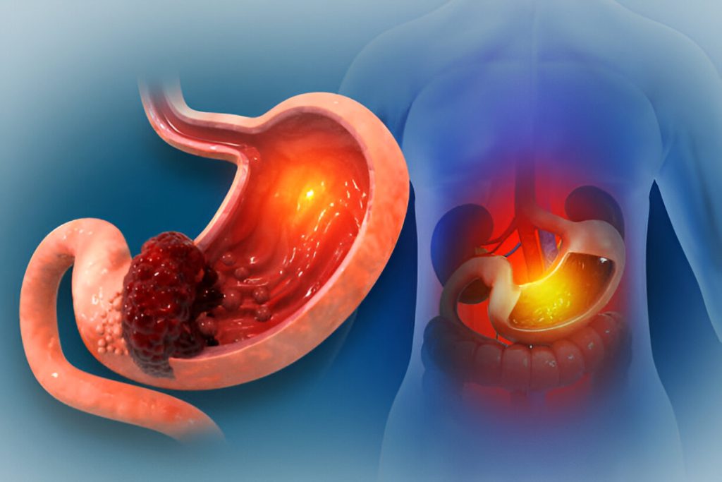
Gastrointestinal Stromal Tumor (GIST) is a type of tumor that develops in the gastrointestinal tract, most commonly in the stomach or small intestine. GISTs arise from specialized cells involved in the movement and coordination of the digestive tract. They can vary considerably in size, behavior, and risk, so accurate diagnosis and individualized management are important.
Some GISTs are discovered incidentally during investigations for other digestive problems, while others may cause symptoms such as abdominal discomfort, gastrointestinal bleeding, anemia, early fullness, nausea, vomiting, or a noticeable abdominal mass.
1. Comprehensive Patient Assessment
Evaluation begins with a detailed consultation with a gastroenterologist. The doctor reviews the patient's symptoms, medical history, previous investigations, medication use, and any history of gastrointestinal disease.
Particular attention may be given to:
Persistent or recurrent abdominal pain
Unexplained gastrointestinal bleeding
Black or tarry stools
Vomiting or nausea
Feeling full after eating a small amount
Unexplained anemia
Loss of appetite
Unintentional weight loss
A suspected abdominal mass
The duration and progression of symptoms are also assessed to determine which investigations may be appropriate.
2. Understanding GIST
GISTs are mesenchymal tumors of the gastrointestinal tract. They are different from the more common cancers that arise from the lining of the digestive tract.
GISTs most frequently occur in the stomach and small intestine, although they can develop elsewhere in the gastrointestinal tract.
Their behavior can range from relatively low-risk tumors to tumors with a significant potential for aggressive growth or spread. Therefore, the size, location, cellular characteristics, and other pathological features are important when determining the risk category and treatment approach.
3. Diagnostic Imaging
Imaging plays an important role in evaluating a suspected GIST.
Depending on the clinical situation, the doctor may recommend:
Contrast-enhanced CT scan
MRI in selected cases
Other imaging investigations when clinically appropriate
Imaging can help determine the location and size of the tumor and assess its relationship with surrounding structures. It may also help evaluate whether there are signs of disease spread.
4. Upper GI Endoscopy
If the suspected tumor is located in the upper gastrointestinal tract, an Upper GI Endoscopy may be performed.
During endoscopy, the gastroenterologist examines the esophagus, stomach, and duodenum using a flexible camera.
A GIST may appear as a subepithelial or submucosal bulge beneath the normal-appearing gastrointestinal lining. Endoscopy allows direct visualization and helps determine whether further assessment is necessary.
5. Endoscopic Ultrasound (EUS)
Endoscopic Ultrasound (EUS) can provide detailed information about a suspected GIST, particularly when the lesion is located in the stomach or upper gastrointestinal tract.
EUS combines endoscopy with ultrasound imaging to evaluate the different layers of the gastrointestinal wall.
It can help assess:
Tumor size
Layer of origin
Internal characteristics
Relationship with nearby structures
Features that may influence further management
When appropriate, EUS can also assist in obtaining tissue for pathological evaluation.
6. Tissue Diagnosis and Biopsy
Not every suspected GIST requires the same type of tissue sampling. The decision to obtain a biopsy depends on the tumor's location, size, appearance, treatment plan, and clinical circumstances.
When tissue confirmation is required, an appropriate biopsy technique may be selected, sometimes with the assistance of EUS.
The tissue is examined by a pathologist to determine the nature of the tumor and to perform relevant immunohistochemical testing.
7. Pathological Evaluation
Pathological assessment is an important part of GIST diagnosis.
The laboratory may evaluate specific markers that support the diagnosis of GIST, including commonly used markers such as KIT (CD117) and DOG1.
The pathologist may also assess features such as:
Tumor cell type
Mitotic activity
Tumor size
Tumor location
Presence of necrosis or other relevant pathological features
These findings help clinicians understand the biological behavior of the tumor and guide treatment planning.
8. Risk Assessment
GIST management is not based on tumor size alone.
The healthcare team considers multiple factors, including:
Tumor size
Tumor location
Mitotic rate
Tumor rupture
Pathological findings
These characteristics help classify the risk of recurrence and guide decisions regarding treatment and follow-up.
9. Multidisciplinary Treatment Planning
Once a GIST has been diagnosed, treatment is planned according to the individual patient's condition.
Depending on the tumor characteristics, management may involve a gastroenterologist, surgeon, medical oncologist, radiologist, pathologist, and other specialists when required.
The treatment plan takes into account whether the tumor is localized, potentially removable, recurrent, or has spread to other parts of the body.
10. Surgical Management
For localized GISTs that are appropriate for removal, surgical resection is an important treatment option.
The aim of surgery is to remove the tumor completely while preserving as much healthy gastrointestinal tissue as reasonably possible.
The exact surgical approach depends on:
Tumor location
Tumor size
Relationship to surrounding structures
Overall health of the patient
Risk of tumor rupture
The surgical team determines the most appropriate approach for each individual case.
11. Targeted Therapy
Some GISTs are driven by specific molecular changes, particularly alterations involving KIT or PDGFRA.
Targeted medicines known as tyrosine kinase inhibitors (TKIs) can be used in appropriate patients. These medicines are designed to interfere with molecular pathways that help certain GIST cells grow.
Imatinib is an important targeted therapy used in many GIST cases, particularly in tumors with specific molecular characteristics. Other targeted therapies may be considered depending on mutation status, treatment response, recurrence, or resistance.
Treatment selection should always be individualized by the treating specialist.
12. Molecular Testing
Molecular testing can provide valuable information about the genetic characteristics of a GIST.
Testing may identify specific mutations, such as changes involving:
KIT
PDGFRA
Other molecular pathways in selected cases
Molecular findings can help guide targeted treatment decisions and may be particularly important when selecting systemic therapy.
13. Management of Advanced or Metastatic GIST
If a GIST has spread to other organs or cannot be completely removed surgically, systemic targeted therapy may become an important part of treatment.
Treatment is selected according to the tumor's molecular characteristics, previous therapies, response to treatment, and overall patient condition.
Some patients may require long-term medical treatment and regular imaging to monitor response.
14. Monitoring Treatment Response
Patients receiving treatment may require regular follow-up and imaging studies.
The purpose of monitoring is to assess:
Tumor response
Tumor progression
Development of new lesions
Treatment-related effects
Need for changes in management
CT or MRI may be used according to the location and clinical situation.
15. Follow-Up After Treatment
Even after successful treatment, regular follow-up may be recommended because some GISTs can recur.
Follow-up schedules depend on the tumor's risk category, treatment received, and individual clinical circumstances.
During follow-up, the healthcare team may review symptoms, perform physical assessments, and arrange appropriate imaging or other investigations.
16. Managing Gastrointestinal Bleeding
Some GISTs can cause gastrointestinal bleeding because the tumor may affect the overlying gastrointestinal lining.
Patients may experience:
Black stools
Vomiting blood
Fatigue
Weakness
Anemia
Gastrointestinal bleeding requires appropriate medical assessment. Depending on its severity, endoscopic, radiological, surgical, or other interventions may be required.
17. Patient Support and Ongoing Care
A diagnosis of GIST can require long-term medical follow-up. Patients may benefit from clear communication between their gastroenterologist, surgeon, oncologist, radiologist, and pathology team.
Ongoing care may include monitoring treatment response, managing side effects, reviewing imaging results, and adjusting treatment when clinically necessary.
When to Seek Medical Evaluation
Patients should seek medical assessment if they experience persistent or unexplained:
Abdominal pain
Gastrointestinal bleeding
Black stools
Vomiting
Unintentional weight loss
Loss of appetite
Early fullness
Unexplained anemia
A new or enlarging abdominal mass
Early evaluation can help identify the underlying cause and determine whether further gastrointestinal investigations are needed.
Comprehensive GIST Care
Our gastroenterology service provides a structured approach to the evaluation of suspected and confirmed gastrointestinal stromal tumors. This may include detailed clinical assessment, endoscopic evaluation, Endoscopic Ultrasound (EUS) when appropriate, imaging, tissue diagnosis, pathological assessment, and coordination with surgical and oncology specialists.
Treatment and follow-up are individualized according to the tumor's location, size, pathological characteristics, molecular profile, and overall clinical condition.
Conclusion
Gastrointestinal Stromal Tumor (GIST) is a distinct type of gastrointestinal tumor that requires accurate diagnosis and individualized management. Because GISTs can behave differently from one another, assessment of tumor location, size, pathology, mitotic activity, molecular characteristics, and other risk factors is essential.
With appropriate diagnostic evaluation, multidisciplinary treatment planning, targeted therapy when indicated, surgical management when appropriate, and regular follow-up, patients can receive comprehensive care throughout the course of their condition...
BEST GASTROENTEROLOGIST IN KARACHI
• Dr AK Lohana Clinic & Endoscopy Services
• Hilal Ahmer Hospital, Clifton
• The Best Doctor in Karachi – Gastroenterologist
• Online Consultation Available
• Book an Appointment
• Call / WhatsApp: 0334-3646307