Esophageal Dilatation
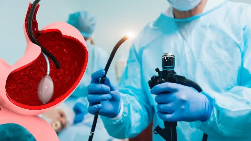
Esophageal Dilatation is an endoscopic procedure used to widen a narrowed or obstructed area of the esophagus, the muscular tube that carries food and liquids from the mouth to the stomach. Esophageal narrowing, also known as an esophageal stricture, can make swallowing difficult and may result from conditions such as acid reflux, scarring, previous inflammation, certain treatments, or other disorders affecting the esophagus.
The main goal of esophageal dilatation is to safely restore a more open passage so that food and liquids can pass more easily from the esophagus into the stomach.
Depending on the patient's condition, dilatation may be performed using a balloon dilator or a bougie dilator. The choice of technique depends on the location, severity, length, and cause of the narrowing, as well as the patient's overall clinical condition.
Step 1: Understanding Esophageal Narrowing
The esophagus normally provides a smooth passage for food and liquids. When part of the esophagus becomes narrowed, swallowing can become difficult.
This narrowing may be associated with:
Difficulty swallowing solid foods
Difficulty swallowing liquids in some cases
A sensation of food getting stuck
Pain or discomfort while swallowing
Regurgitation of food
Recurrent episodes of choking or coughing while eating
Unintentional weight loss when swallowing difficulty affects food intake
An esophageal narrowing can have different underlying causes, so appropriate evaluation is important before treatment.
Step 2: Common Causes of Esophageal Stricture
Esophageal narrowing can occur for several reasons.
Common causes may include:
Acid Reflux and GERD
Long-term exposure of the esophagus to stomach acid can cause inflammation and, in some patients, scarring. This scar tissue can gradually narrow the esophageal passage.
Peptic or Inflammatory Injury
Repeated inflammation or ulceration of the esophageal lining can result in scar formation and narrowing.
Eosinophilic Esophagitis
This inflammatory condition can cause swelling, fibrosis, and narrowing of the esophagus in some patients.
Previous Procedures or Treatments
Certain medical procedures or treatments involving the esophagus or surrounding area may contribute to scarring and narrowing.
Other Structural Causes
Benign strictures, rings, webs, and other abnormalities can also interfere with the normal passage of food.
In some patients, narrowing may also be related to a tumor or other serious underlying condition. Therefore, proper assessment is essential before or during treatment.
Step 3: Medical Assessment Before Dilatation
Before performing esophageal dilatation, the gastroenterologist reviews the patient's symptoms and medical history.
The assessment may include:
Duration and severity of swallowing difficulty
Whether difficulty occurs with solids, liquids, or both
History of GERD or chronic acidity
Previous esophageal disease
Previous endoscopic procedures
Previous surgery or radiation treatment
Medication history
Weight loss or nutritional problems
Previous biopsies or imaging results
The doctor may also assess whether the patient has symptoms that require urgent or additional investigation.
Step 4: Diagnostic Endoscopy
In many patients, an Upper GI Endoscopy (EGD/Gastroscopy) is performed to directly examine the esophagus.
The endoscope allows the gastroenterologist to visualize:
Mouth → Esophagus → Stomach → Duodenum
During the examination, the doctor can identify the narrowed area, assess its appearance, determine how severe it is, and look for inflammation, ulcers, rings, webs, or other abnormalities.
When appropriate, biopsies may also be taken to investigate the underlying cause of the narrowing.
Step 5: Preparing the Patient
Proper preparation is important for safe and effective esophageal dilatation.
The patient is generally instructed to:
Remain fasting for the required period before the procedure
Follow the doctor's instructions regarding food and fluids
Inform the medical team about all medicines being taken
Inform the doctor about blood-thinning medicines
Report any known medication or anesthesia allergies
Provide information about previous medical conditions and procedures
The exact preparation plan depends on the patient's condition and the sedation or anesthesia protocol being used.
Step 6: Sedation and Monitoring
Esophageal dilatation is commonly performed with appropriate sedation or anesthesia to improve comfort and allow the procedure to be performed safely.
During the procedure, the medical team may monitor:
Blood pressure
Heart rate
Oxygen saturation
Breathing
Overall clinical condition
The level of sedation is selected according to the patient's needs, procedure complexity, and clinical circumstances.
Step 7: Introduction of the Endoscope
The gastroenterologist carefully introduces the endoscope through the mouth and into the esophagus.
The camera at the tip of the endoscope provides a clear view of the esophageal lining and allows the doctor to identify the exact location of the narrowing.
The narrowed segment is evaluated before dilatation is performed.
Step 8: Identifying and Assessing the Narrowed Area
The gastroenterologist carefully examines the stricture to determine:
Its location
Length
Severity
Diameter of the narrowed passage
Appearance of the surrounding tissue
Presence of inflammation or ulceration
Presence of any suspicious lesion
This assessment helps the doctor select the most appropriate dilatation technique and size.
Step 9: Balloon Esophageal Dilatation
One commonly used technique is Balloon Esophageal Dilatation.
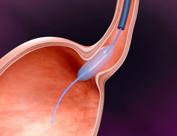
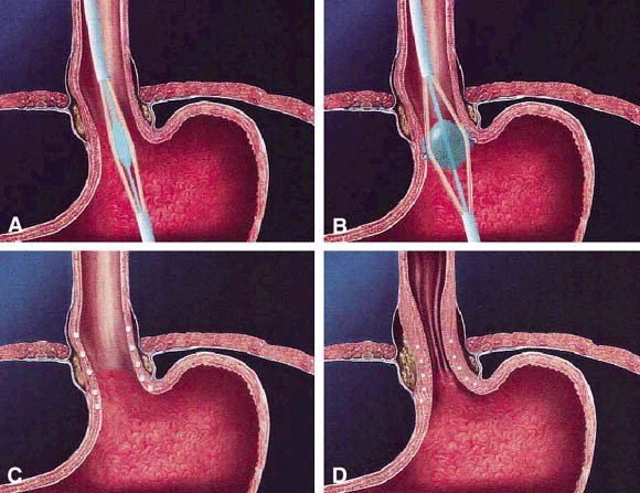
A specially designed deflated balloon catheter is positioned across the narrowed segment of the esophagus.
The balloon is then gradually inflated in a controlled manner.
As it expands, the balloon applies gentle radial pressure to the narrowed area and widens the esophageal passage.
The doctor carefully controls:
Balloon position → Inflation → Pressure → Duration → Deflation
The exact technique depends on the type and severity of the stricture.
Step 10: Bougie Dilatation
In selected cases, a bougie dilator may be used instead of a balloon.
A bougie is a smooth, flexible dilating instrument that is gradually passed through the narrowed area to increase the diameter of the esophageal passage.
The choice between balloon and bougie dilatation depends on the location and characteristics of the narrowing, the doctor's experience, and the patient's individual clinical situation.
Step 11: Gradual and Controlled Widening
Esophageal dilatation is performed carefully and progressively.
The objective is to improve the diameter of the narrowed segment while minimizing unnecessary trauma to the esophageal wall.
In some patients, more than one dilatation session may be necessary, particularly when the narrowing is severe, long-standing, or recurrent.
The treatment plan is individualized according to the patient's symptoms and response.
Step 12: Biopsy When Clinically Indicated
If the endoscopic appearance suggests inflammation, eosinophilic esophagitis, Barrett's esophagus, a suspicious lesion, or another underlying condition, the gastroenterologist may obtain tissue samples.
A biopsy can help determine the reason for the narrowing and guide further treatment.
Taking a biopsy does not automatically mean that cancer is suspected; tissue sampling is often performed to establish an accurate diagnosis.
Step 13: Assessing the Result During the Procedure
After dilatation, the gastroenterologist reassesses the treated area.
The doctor evaluates whether:
The passage has widened adequately
The endoscope can pass more easily
The mucosal appearance is satisfactory
Further dilatation may be required
Any additional abnormality needs investigation or treatment
The procedure is stopped or modified according to the patient's condition and the findings.
Step 14: Causes That May Require Additional Treatment
Esophageal dilatation treats the narrowing, but it may not by itself eliminate the underlying cause.
For example, when strictures are associated with acid reflux, ongoing management of GERD may be necessary to reduce the risk of recurrence.
Depending on the underlying condition, additional management may involve:
Acid-suppressive therapy
Treatment of inflammatory esophageal disease
Management of eosinophilic esophagitis
Follow-up endoscopy
Repeat dilatation
Biopsy and pathology assessment
Treatment of another underlying esophageal disorder
A comprehensive treatment plan is therefore important for long-term results.
Step 15: Benefits of Esophageal Dilatation
Esophageal dilatation may provide significant improvement in swallowing symptoms.
Potential benefits include:
Easier swallowing
Improved passage of food and liquids
Reduced sensation of food sticking
Improved ability to maintain adequate nutrition
Relief from symptoms caused by esophageal narrowing
Improved quality of life
The degree and duration of improvement depend on the underlying cause and severity of the stricture.
Step 16: Repeat Dilatation and Long-Term Management
Some esophageal strictures may recur after treatment.
Patients with recurrent narrowing may require additional dilatation sessions at intervals determined by their gastroenterologist.
Repeated treatment may be necessary when:
The stricture is severe
The narrowing is long-standing
Scar tissue reforms
The underlying disease remains active
Symptoms return after an initial improvement
Regular follow-up helps detect recurrence and allows treatment to be adjusted appropriately.
Step 17: Recovery After the Procedure
After the procedure, the patient is monitored while recovering from sedation or anesthesia.
Some patients may temporarily experience:
Mild throat discomfort
Mild chest discomfort
Temporary bloating
Mild difficulty swallowing immediately after the procedure
These symptoms may improve over a short period.
The medical team provides individualized instructions regarding when the patient can resume eating, drinking, and normal activities.
Step 18: Diet After Esophageal Dilatation
Dietary instructions after dilatation depend on the patient's condition and the doctor's recommendations.
Patients may initially be advised to start with fluids or soft foods before gradually returning to their normal diet.
The exact diet depends on:
The extent of dilatation
The underlying condition
Whether a biopsy or additional treatment was performed
The patient's swallowing ability
The gastroenterologist's assessment
Patients should follow the specific post-procedure instructions provided by their healthcare team.
Step 19: Possible Risks and Complications
Esophageal dilatation is generally performed by trained specialists and is considered a well-established procedure, but like any medical intervention, it carries potential risks.
Possible complications may include:
Temporary chest or throat discomfort
Minor bleeding
Infection, which is uncommon
Adverse reaction to sedation or anesthesia
Recurrent or persistent narrowing
Esophageal perforation, a rare but potentially serious complication
The individual risk depends on the cause and severity of the narrowing, the technique used, and the patient's overall health.
Step 20: Warning Signs After the Procedure
Patients should follow their discharge instructions carefully and contact their healthcare provider if concerning symptoms develop.
Urgent medical assessment may be required in the presence of:
Severe or worsening chest pain
Severe abdominal pain
Difficulty breathing
Persistent vomiting
Significant or ongoing bleeding
Fever
Sudden deterioration in overall condition
These symptoms should be evaluated promptly because they may indicate a complication requiring immediate treatment.
Step 21: Who May Benefit From Esophageal Dilatation?
Esophageal dilatation may be considered for appropriately selected patients with:
Benign esophageal strictures
Difficulty swallowing caused by narrowing
Peptic strictures associated with chronic reflux
Esophageal rings or webs
Selected inflammatory esophageal conditions
Recurrent esophageal narrowing
Other structural causes of esophageal obstruction
The procedure is not appropriate for every swallowing problem, and a proper diagnosis is required before treatment.
Step 22: Why Specialist Evaluation Is Important
Difficulty swallowing should not be ignored, particularly when it is persistent or progressively worsening.
A gastroenterologist can determine whether the symptoms are caused by:
Esophageal narrowing → Inflammation → Motility disorder → Reflux disease → Structural abnormality → or another underlying condition
Upper GI Endoscopy and other investigations may be recommended when clinically appropriate.
Early diagnosis helps ensure that the underlying condition is identified and treated alongside the narrowing itself.
Frequently Asked Questions About Esophageal Dilatation
What is Esophageal Dilatation?
Esophageal dilatation is a procedure used to widen a narrowed area of the esophagus and improve the passage of food and liquids.
Why does the esophagus become narrow?
Common causes include scarring from chronic acid reflux, inflammation, eosinophilic esophagitis, rings or webs, and other structural or medical conditions.
Is esophageal dilatation performed through an endoscope?
In many cases, yes. The gastroenterologist uses an endoscope to visualize the esophagus and guide the dilatation procedure.
What is balloon dilatation?
Balloon dilatation involves positioning a deflated balloon across the narrowed area and carefully inflating it to widen the esophageal passage.
Is one session always enough?
Not necessarily. Some patients may require more than one session depending on the severity, length, and cause of the narrowing and whether it recurs.
Does esophageal dilatation treat acid reflux?
Dilatation treats the narrowing caused by reflux-related scarring, but the underlying GERD may still require ongoing medical treatment to reduce the risk of recurrence.
Can a biopsy be performed during the procedure?
Yes. When clinically indicated, tissue samples can be collected during endoscopy to investigate inflammation or other abnormalities.
Is esophageal dilatation painful?
The procedure is generally performed with appropriate sedation or anesthesia to improve patient comfort. Some temporary discomfort may occur afterward.
How long does recovery take?
Recovery varies depending on the procedure and sedation used. Many patients can resume normal activities relatively quickly, according to their doctor's instructions.
Conclusion
Esophageal Dilatation is an established endoscopic procedure for treating narrowing of the esophagus and improving swallowing. Using controlled balloon or bougie techniques, the gastroenterologist carefully widens the narrowed segment to restore a better passage for food and liquids.
Successful management requires more than simply widening the esophagus. Identifying and treating the underlying cause of the narrowing is essential to reduce recurrence and achieve better long-term results.
Patients experiencing persistent difficulty swallowing, recurrent food impaction, painful swallowing, unexplained weight loss, or a sensation that food is becoming stuck in the chest should seek timely evaluation by a qualified gastroenterologist.
Early assessment, accurate diagnosis, appropriate dilatation, and continued follow-up can help improve swallowing function and overall digestive health.